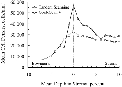
Figure 6.
Mean cell density in anterior stroma recorded by the ConfoScan 4 and Tandem Scanning confocal microscopes, aligned on the frame with maximum cell density. Maximum cell density was higher and the peak was sharper in scans by the Tandem Scanning microscope, characteristics of its thinner depth of field and the thin, high-density layer of cells in the anterior boundary of the stroma. Cell nuclei appeared in images of the ConfoScan 4 (and increased the apparent density) at a greater distance from their location because of the greater depth of field.

