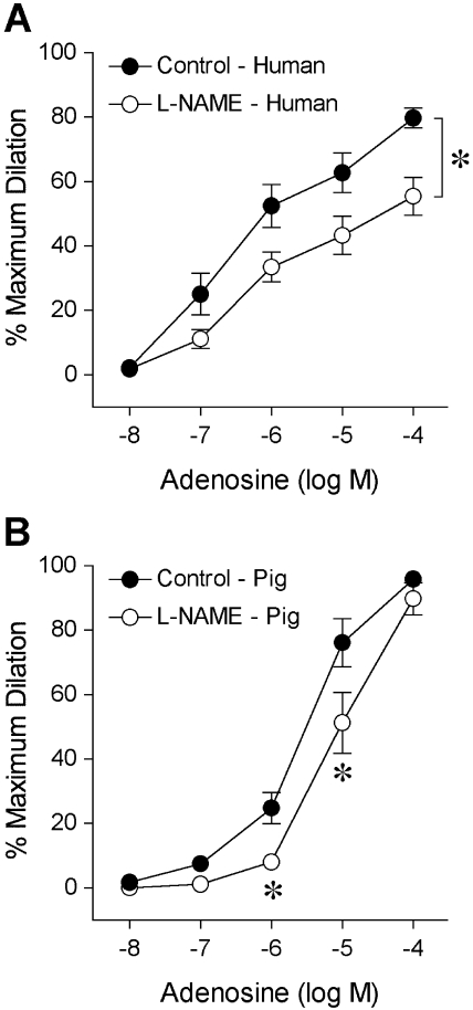
Figure 3.
Vasodilator response of isolated and pressurized human (A) and porcine (B) retinal arterioles to adenosine. The control human (resting diameter, 44 ± 4 μm; maximum diameter, 65 ± 4 μm; n = 9) and porcine (resting diameter, 45 ± 4 μm; maximum diameter, 82 ± 5 μm; n = 8) retinal arterioles dilated dose dependently to adenosine. In the presence of L-NAME (10 μM), the adenosine-induced dilation of both human (resting diameter, 43 ± 4 μm; maximum diameter, 65 ± 4 μm; n = 9) and porcine (resting diameter, 44 ± 4 μm; maximum diameter, 82 ± 5 μm; n = 8) vessels was significantly attenuated. *P < 0.05 versus control.

