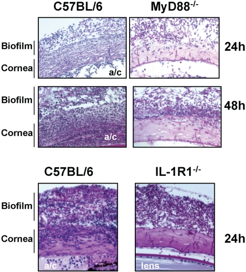
Figure 4.
Histopathology of Fusarium keratitis in MyD88−/− and IL-1R1−/− mice. C57BL/6, MyD88−/−, and IL-1R1−/− mice were given a 1-mm diameter corneal abrasion and a contact lens with adherent Fusarium biofilm was placed on the corneal surface, as described in the legend to Figure 1. After 24 or 48 hours, the eyes were processed for histology and stained with PAS. Representative images of five mice per group are shown. Note that cellular infiltration of the corneal stroma was impaired in the MyD88−/− and IL-1R1−/− mice compared with that in the C57BL/6 mice. a/c, anterior chamber.

