Figure 5.
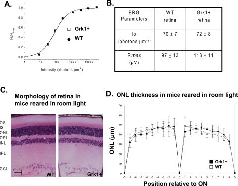
Physiology and morphology of Grk1+ retina. (A) Fractional a-wave amplitude of transretinal ERG recordings as a function of flash intensity from WT (n = 8) and Grk1+ (n = 6) retinas. Data were fit with the equation in the text. (B) Physiological parameters obtained from WT and Grk1+ retinas. Io is the flash intensity that gives the half-maximum response. Rmax is the maximum amplitude of a-wave. Data are expressed as the mean ± SEM. (C) Structure of Grk1+ and WT retinas were viewed by light microscopy. Sections (3 μm) of plastic-embedded retina were cut along the vertical meridian of the globe at the level of the optic nerve and stained with hematoxylin and eosin. OS, outer segment; IS, inner segment; ONL, outer nuclear layer; OPL, outer plexiform layer; INL, inner nuclear layer; IPL, inner plexiform layer; GCL, ganglion cell layer; ON, optic nerve; O, ora serrata. (D) Comparison of Grk1+ and WT ONL thicknesses in micrometers at nine equidistant positions along the superior and inferior hemiretina (+0–9 superior retina, −9–0 inferior retina). Data are expressed as the mean ± SD (n = 8 WT and 8 Grk1+). Scale bar, 50 μm.

