Figure 7.
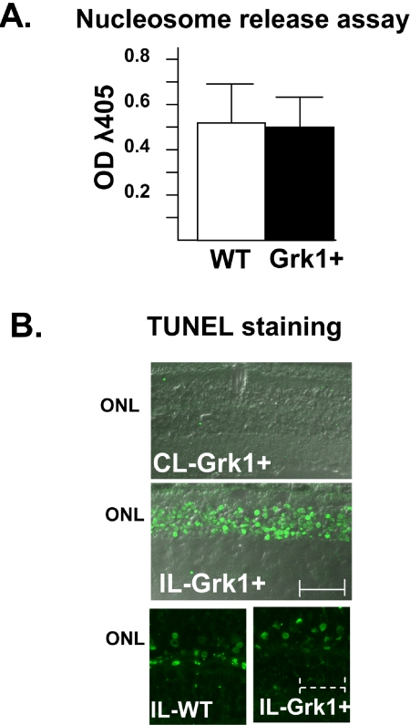
Apoptosis in Grk1+ and WT retinas. (A) Nucleosome release was measured in Grk1+ and WT retinas using immunoassay. Litter, sex, Rpe65 genotype (Leu450Met), and pigment-matched mice were exposed to either cyclic light (CL, only GRK+) or intense light (10,000 lux) for 12 hours. Diluted retinal lysate from individual eyes (20 μL) was examined for the presence of immunoreactive nucleosomes by using sandwich ELISA in microtiter well plates. Absorbance at λ405 nm (OD) was measured on a plate reader (n = 8 for both Grk1+ and WT). (B) TUNEL staining of Grk1+ and WT eyes was performed after exposure of the mice to ambient cyclic light (CL, 60 lux) or intense light (IL,10,000 lux) after 12 to 24 hours of dark adaptation. Scale bar: solid, 25 μm; dashed, 7 μm.

