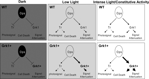
Figure 8.
A model for Grk1-mediated photoreceptor cell death. Phototransduction pathways in dark, low, and intense light in model WT and Grk1+ photoreceptors are illustrated. In the absence of light, the light-dependent pathways including transducin-dependent visual signaling and Grk1-mediated deactivation are both quiescent, regardless of the levels of Grk1. In low light, both light-dependent pathways become stimulated with increased rhodopsin flow through both activation and deactivation channels. In this state, biased flow toward the deactivation pathway has little impact on photoreceptor integrity and may actually be protective. In intense light, the differential flow of signal through the Grk1-mediated deactivation pathway becomes a liability acting as a transducin-independent cell death pathway. Dashed arrows: subnormal flux through a pathway below that expected of the WT levels. Progressively bolder solid arrows: increased flux over those in the WT state.

