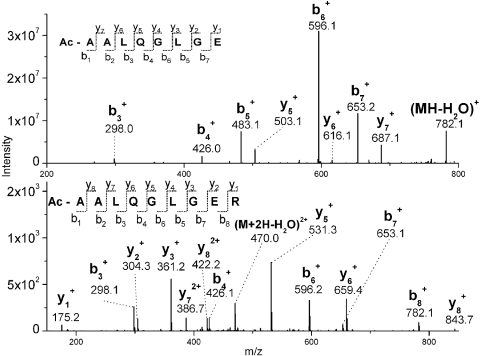
Figure 5.
Tandem mass spectra of the Glu C peptide acetyl-AALQGLGE (40–47) (top) and the tryptic peptide acetyl-AALQGLGER (40–48) (bottom) of filensin are plotted. Tandem mass spectra are labeled with the predicted b- and y-ions. All b-ions are shifted in mass by 42 Da from their expected m/z values.

