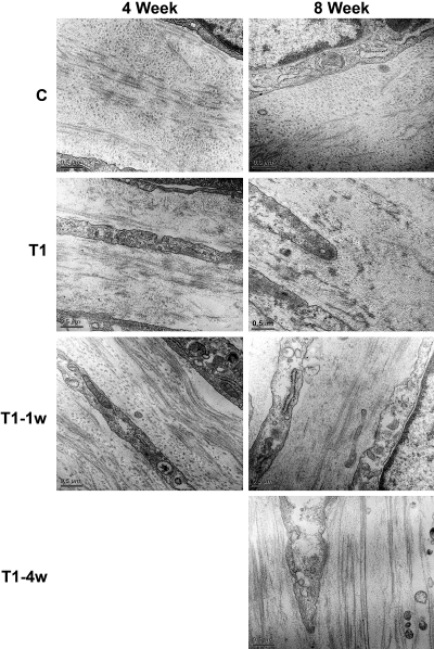
Figure 2.
High-magnification TEM (31,000×) showing cell–matrix interaction and matrix condition. The fibril orientation changed direction more than once in all the conditions at both 4 and 8 weeks; however, by 8 weeks, T1 fibril integrity appeared to be decreasing. Of interest, at both the 4- and 8-week time points, the fibrils in T1–1w and T1–4w appeared to be longer than those in C. Bar, 0.5 μm.

