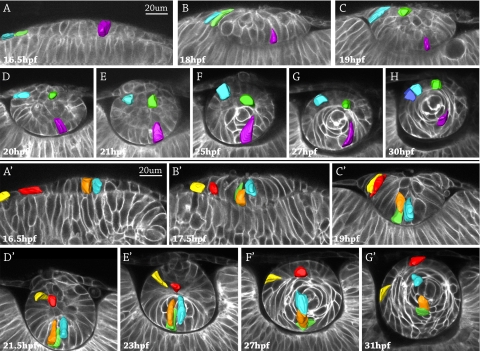
Figure 1.
Cell fate tracking during lens development in two representative zebrafish embryos. Image stacks were acquired every 30 minutes, and representative images are shown. The surface ectoderm is oriented at the top of each image, and the retina is at the bottom. Three pseudocolored cells were followed from the lens placode stage at 16.5 hpf to obvious lens differentiation into an anterior epithelium and fiber cells at 30 hpf (A–H). Five pseudocolored cells in a different embryo were followed from 16.5 hpf to 31 hpf (A′–G′). The violet cell (A–H) and the orange and blue cells in (A′–G′) started in the central placode, moved to the posterior lens mass (where the orange cell divided into two cells, orange and green), and elongated as primary fibers in the embryonic lens nucleus. The blue and green cells (A–H) and the red and yellow cells (A′–G′) originated in the peripheral placode, migrated to the anterior lens mass, and became part of the anterior epithelium.

