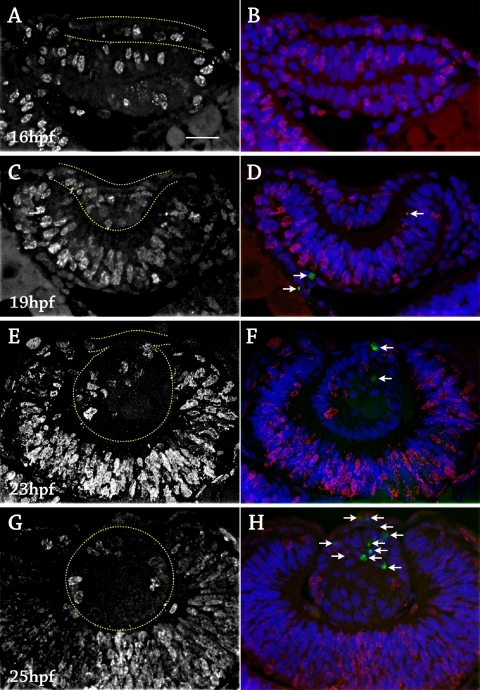
Figure 3.
Cell birth and death in the zebrafish lens, 16 to 25 hpf. Fixed lens sections stained with anti-BrdU for proliferation (red), TUNEL for apoptosis (green, white arrows), and DAPI to label cell nuclei (blue). Six to 12 embryos were examined at each time point. (A) 16 hpf, anti-BrdU alone. The lens placode was outlined in yellow. Proliferation occurred throughout the placode and retina. (B) Same section as in A with DAPI and TUNEL. No apoptotic cells were detected. (C) 19 hpf, anti-BrdU alone. The lens mass was outlined in yellow. Proliferation occurred throughout the lens mass and retina. (D) Same section as in C with DAPI and TUNEL. A few apoptotic cells were detected in the retina (arrows). (E) 23 hpf, anti-BrdU alone. The delaminating lens mass was outlined in yellow. Proliferation was mainly in the anterior lens mass. (F) Same section as in E with DAPI and TUNEL. Two apoptotic cells were visible in the anterior lens mass (arrows). (G) 25 hpf, anti-BrdU alone. The lens was outlined in yellow. Proliferation was restricted to the lens epithelium. (H) Same section as in G with DAPI and TUNEL. Multiple apoptotic cells were detected in the anterior lens mass and cornea after separation (arrows). Scale bar, 20 μm.

