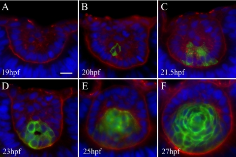
Figure 5.
Zl-1 antibody staining primary lens fiber cells. Sections were labeled with Zl-1 (green), wheat germ agglutinin (red), DAPI (blue). Six to 12 embryos were examined at each time point. (A) The 19 hpf lens contained no Zl-1 staining. (B) At 20 hpf, one cell in the posterior lens mass stained with Zl-1. Primary fibers cell differentiation begins. (C, D) At 21.5 hpf and 23 hpf, increasing numbers of elongating primary fiber cells at the posterior lens mass stained with Zl-1. (E) At 25 hpf after separation of the lens and cornea, all cells in the posterior lens were Zl-1 positive, and cells in the anterior lens, which were organizing into the anterior epithelium, did not stain. (F) At 27 hpf, formation of a single layer of anterior epithelium wrapping around the lens was nearly complete. All primary fibers were Zl-1 positive.

