Figure 4.
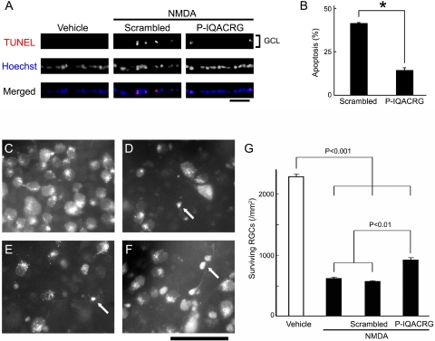
P-IQACRG protects RGCs from apoptosis after intravitreal NMDA injection. (A) NMDA (15 nmol) and glycine (10 nmol) were injected intravitreally along with 150 pmol P-IQACRG or scrambled (control) peptide. Eight hours later, retinal sections were subjected to TUNEL staining (red in merged image) to assess apoptosis and were counterstained with Hoechst 33342 to label cell nuclei (blue); colabeling appears violet. Injection of NMDA led to apoptosis in the GCL, whereas vehicle (glycine alone) did not. Administration of P-IQACRG but not the scrambled peptide protected the GCL from apoptosis. Scale bar, 50 μm. (B) Quantitative assessment of number of TUNEL-positive nuclei in the GCL revealed that P-IQACRG inhibited apoptosis induced by NMDA (*P < 0.001, Student's t-test). Data are mean ± SEM (n = 5–6 for each group). (C–F) Representative photomicrographs of aminostilbamidine-labeled RGCs in retinal flat-mounts 24 hours after intravitreal injection of vehicle (glycine alone; n = 4; C), NMDA plus glycine (n = 5; D), NMDA plus glycine treated with scrambled peptide (n = 9; E), or NMDA plus glycine treated with P-IQACRG (n = 10; F). NMDA injection resulted in fewer RGCs and pyknotic-appearing somas in many of the remaining cells (arrows). Scale bar, 50 μm. (G) Surviving RGCs were counted in retinal flat-mounts and were expressed as RGC density per square millimeter. Each bar represents mean ± SEM. Statistical significance was assessed with a one-way ANOVA followed by Fisher's PLSD multiple comparisons test.

