Figure 5.
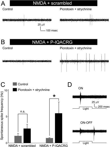
Electrophysiological activity of surviving RGCs. (A, B) Retinal flat-mounts were prepared for electrophysiological analysis 24 hours after intravitreal injection of NMDA (15 nmol) plus glycine (10 nmol) with either scrambled peptide or P-IQACRG (150 pmol). Ex vivo retinal recordings were performed on MEAs initially in normal extracellular solution (control, left) and then after the addition of picrotoxin (100 μM) and strychnine (10 μM) to suppress inhibitory input (right). RGCs exhibited spontaneous spiking activity, which increased in the presence of picrotoxin and strychnine, particularly in retinas treated with P-IQACRG. (C) Spontaneous spike frequency obtained from multiple electrodes was averaged for each retina and compared (n = 6 retinas for each treatment). P-IQACRG-treated retinas exhibited a statistically significant increase in spike frequency in the presence of picrotoxin and strychnine (*P < 0.05; n.s., not significant). (D) Retinal responses to full-field illumination (1 second in duration). Surviving RGCs in P-IQACRG-treated retinas displayed light-evoked responses. These traces represent recordings in medium lacking picrotoxin and strychnine and illustrate an ON response (top) and an ON-OFF response (bottom).

