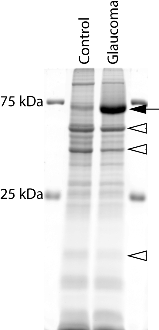
Figure 1.
Identification of a 70 kDa protein overexpressed in the retina of monkeys with experimental glaucoma. Total protein dye (Deep Purple; GE Healthcare)-stained gel of retinal proteins from the control (C) and glaucoma (G) eye of the same monkey (OHT 46; inferior 40°–45°) separated by SDS-PAGE. Although equal amounts of proteins (20 μg) were loaded in each lane, the protein content near 70 kDa was obviously greater in the glaucoma retina (lane 2) than in the control retina. This same pattern of labeling is seen in six of the monkeys studied. Only five to six prominent protein bands were detected since the amount of protein loaded in each well is 1.5–6 times lower than that in most studies. Filled arrow: increased 70 kDa protein band; open arrowheads: decreased proteins.

