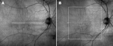
Figure 1.
For Cirrus HD-OCT, the standard macular imaging protocol in the Doheny Ophthalmic Imaging Unit consists of the 5 Line Raster (A) and the Macular Cube (B) scanning protocols. The Macular Cube protocol consists of 128 horizontally oriented B-scans, each 6 mm in length and composed of 512 equally spaced transverse sampled locations.

