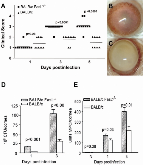
Figure 1.
Disease in BALB/c FasL−/− compared with WT mice. (A) Corneal disease was graded at 1, 3, and 5 days p.i. (n = 5/group/time). BALB/c FasL−/− compared with WT mice had worsened disease at 3 and 5 days p.i. (P < 0.0001). (B, C) Photographs taken with a slit lamp. Eyes from BALB/c WT (B) and FasL−/− mice (C) are shown at 5 days p.i. Original magnification, 15×. (D) Viable bacterial plate counts. BALB/c FasL−/− compared with WT mice (n = 5/group/time) had significantly more bacteria at 1 and 3 days p.i. (P < 0.001). Results are reported as 105 CFU/cornea ± SEM. (E) PMNs per cornea (mean ± SEM; n = 5/group/time) determined by MPO assay. More PMNs were detected in the corneas of BALB/c FasL−/− than WT mice at 1 (P = 0.03) and 3 (P = 0.01) days p.i.

