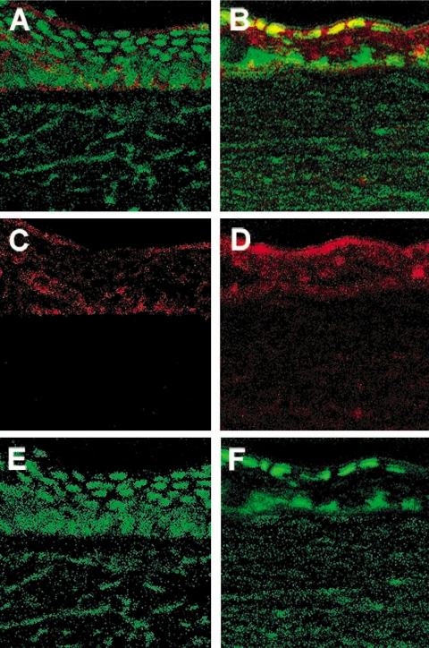
Figure 5.
Immunostaining for Bcl-2 in the infected BALB/c FasL−/− (B, D, F) compared with WT (A, C, E) cornea at 1 day p.i. (n = 3/group/time/test). More intense Bcl-2 staining (red) was detected in FasL−/− compared with WT mouse corneal epithelium (top, merged image; middle, Bcl-2 staining alone). SYTOX green nuclear label (E, F) appeared similar to substitution of the primary antibody with rabbit IgG. Original magnification, 280×.

