
Figure 1.
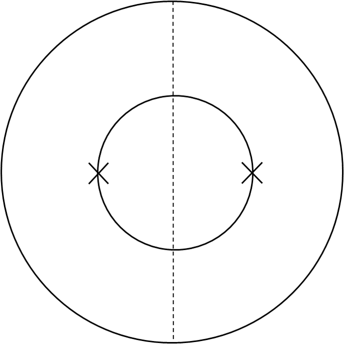
Schematic picture illustrating the suture placement method used to compare the nasal and temporal distributions of vessels. Two sutures were placed at the 3 o'clock and 9 o'clock positions of the cornea, respectively. Outer circle: limbus. Inner circle: demarcation of the trephine. Dashed line: demarcation between the nasal and the temporal sides.