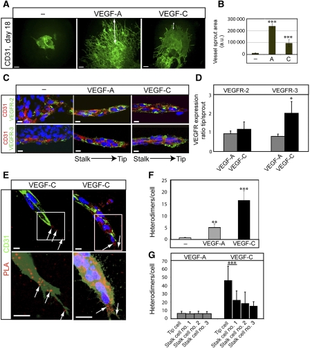
Figure 5.
Heterodimers in angiogenic sprouts. (A) Angiogenic sprouting in response to VEGFA or VEGFC. EBs were cultured in 3D collagen matrix in the presence of VEGFA or VEGFC. Microphotographs were taken at day 18 on whole-mount fixed, CD31-immunostained samples. Scale bar=300 μm. (B) Quantification of total vascular area from the data in (A) based on five EBs per condition and expressed as fold induction ±s.d. (C) Expression of VEGFR2 and VEGFR3 in angiogenic sprouts. Immunostaining for VEGFR2 (green; upper panels) and VEGFR3 (green; lower panels) on CD31-positive (red) angiogenic sprouts in 3D EBs treated with VEGFA or VEGFC. The orientation of the tip versus stalk is indicated. Scale bar=10 μm. (D) Quantification of VEGFR2 and VEGFR3 expression in the tip cell region compared with entire angiogenic sprouts. n=4. (E) Location of heterodimers on VEGFC-induced angiogenic sprouts. Red spots represent PLA reactions in tip cells. Panels show PLA spots in CD31-positive angiogenic tip cell regions. Lower panels show saturation of CD31-positivity to better visualize the filopodia. Note that PLA spots are located on filopodia extending ahead of the tip cell. Scale bar=10 μm. (F) Quantification of in situ PLA detecting VEGFR2/-3 heterodimers in angiogenic sprouts in response to VEGFA or VEGFC in 3D EB cultures. n=8. (G) Quantification of heterodimers in tip and stalk cells. VEGFA-treated angiogenic sprouts contained heterodimers evenly distributed over the sprouts, whereas VEGFC-treated angiogenic sprouts showed accumulation of heterodimers in tip cells. n=8. Asterisks in panels B, D, F–G indicate degree of significance (*P<0.05, **P<0.01, ***P<0.001).

