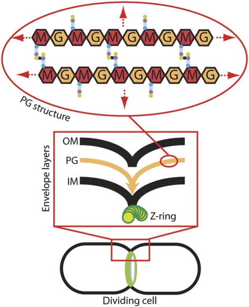
Figure 1.
Coordinated envelope constriction in gram-negative bacteria. Diagram of a dividing cell with an assembled cytokinetic ring apparatus (green). The box contains a close-up diagram of the division site highlighting the coordinated constriction of the envelope layers: OM, outer membrane; PG, peptidoglycan; IM, inner membrane; Z-ring, FtsZ cytoskeletal ring. The oval contains a diagram of the PG chemical structure: M, N-acetylmuramic acid; G, N-acetylglucosamine. Coloured dots represent the attached peptides. The PG structure continues in all directions to envelop the cell (red arrows).

