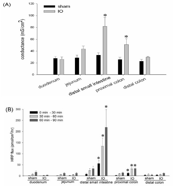
Figure 3.
IO induced epithelial permeability rise in the distal small intestine and proximal colon. Various bowel segments in sham and IO rats were mounted on Ussing chambers for the measurement of electrical conductance (Panel A) and mucosal-to-serosal flux of HRP (Panel B). The rate of HPR flux was determined at different time points after luminal addition of the probe: 0 to 30, 30 to 60, and 60 to 90 min. n = 5-7/group. *p < 0.05 vs. respective sham groups.

