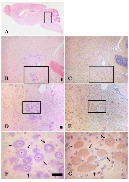
Fig. 3.
Colocalization of synemin transcript and tryptophan hydroxylase-1 protein (TPH-1) in brain sagittal sections. Panels A, B, D, and F are in situ experiments hybridized with the synemin probe-3, whereas panels C, E, and G are mirror sections of panels B, D, and F and are immunostained with an anti-tryptophan hydroxylase-1 antibody. Light purple designates in situ positive structures whereas orange indicates immunostaining with the anti-TPH-1 antibody. Panel A shows a whole sagittal brain where signals with probe-3 are seen inside the square. Panels B, D, and F are magnified of squares in panels A, B, and D, respectively. Squares in B and C show the same area as well as those in D and E. Arrows in F and G indicate the same neurons. Panels B and C are enlarged 50X, D and E are 100X, and F and G are 400X. Bar, 50μm

