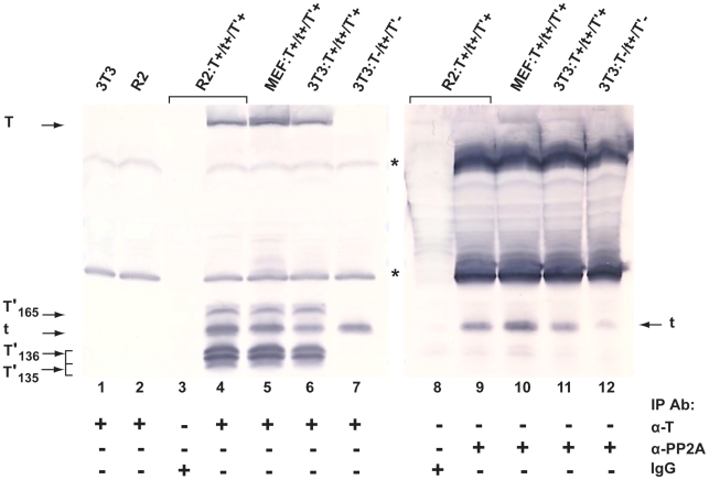
Figure 1. Wild type tAg interacts with cellular phosphatase PP2A in cells expressing JCV early proteins.
JCV early proteins, expressed in Rat 2 (R2) or MEF cells transformed with pSR:T+/t+/T'+ or in G418-selected 3T3 cells transfected with pSR:T+/t+/T'+ (encodes all 5 JCV early proteins) or pSR:T−/t+/T'− (encodes JCV tAg only) were incubated with anti-T monoclonal antibody PAb 962 (α-T; lanes 4–7) or anti-PP2A antibody (α-PP2A; lanes 9–12). The amount of total cell protein subjected to IP in lanes 9–12 was four times that employed in the corresponding samples in lanes 4–7. Immunoprecipitated proteins were separated on a 20% SDS-polyacrylamide gel, and WB analysis was performed using a cocktail of anti-T monoclonal antibodies to detect JCV early proteins either expressed in the different cell lines (lanes 4–7) or expressed and bound to PP2A (lanes 9–12). Untransfected 3T3 and Rat 2 cells were included as negative controls (no JCV T proteins are present; lanes 1, 2), and α-mouse IgG was used in the IP step with the R2:T+/t+/T'+ cell extract to test for non-specific binding (lanes 3, 8). The asterisks denote antibody light and heavy chains. This figure represents proteins electrophoresed on a single gel and transferred to a membrane, which was then cut in half and each half developed for different lengths of time.

