Figure 1.
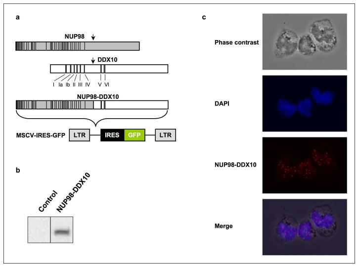
Nuclear expression of NUP98-DDX10 in primary human CD34+ cells. (a) Schematic representation of NUP98, DDX10 and NUP98-DDX10. Vertical lines in the NUP98 portion indicate the locations of FG repeats. The locations of conserved helicase motifs in DDX10 are indicated with Roman numerals. (b) Immunoblotting with anti HA antibody shows NUP98-DDX10 expression in retrovirally transduced primary human CD34+ cells. (c) CD34+ cells retrovirally transduced with HA-NUP98-DDX10 were immunostained with anti-HA antibody and Alexa Fluor 647-conjugated anti-mouse IgG secondary antibody. The corresponding phase contrast image, nuclear counterstain with DAPI, and the merged images are shown. Images were viewed using a Nikon Eclipse 80i microscope with a Nikon 40X, 0.75 numerical aperture CFI Plan Fluor DLL objective and were acquired with a Nikon Coolsnap ES camera using MetaMorph 6.3r2 software with pseudo-coloring. The merged image was obtained by superimposing the 3 images using Photoshop CS4 software.

