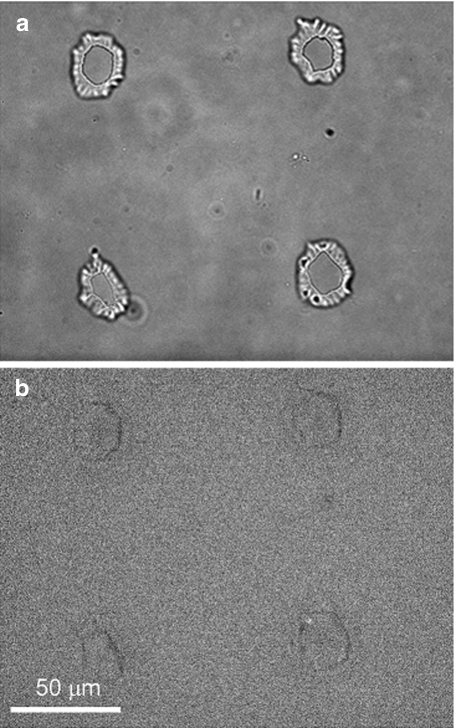
Fig. 1.
a Phase-contrast image of four microarray chambers, each roughly 3 pl in volume. The upper left chamber and both lower chambers initially contained a single V. fischeri cell. The upper right chamber remained empty. The irregular light and dark patterns surrounding each chamber are features of the PDMS walls; the live cells are not readily visible in this image. b Luminescence image of the same chambers after 4.5 h. A region of bioluminescence is visible in the lower right chamber, signaling activation of the lux genes

