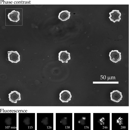
Fig. 2.
Upper: Phase-contrast image of nine chambers (volume 3.4 ± 0.6 pl) containing pAC-LuxGfp E. coli. Cells appear as dark rodlike shapes near the centers of the two lower chambers in the right hand column. The white box indicates the chamber whose fluorescence is imaged in the lower panel. Lower: GFP fluorescence images for the chamber indicated above, collected at various times (with 10-s exposures) after trapping of a single cell. The LB medium gives a weak fluorescent background, but the abrupt onset of GFP fluorescence is apparent at ~120 min

