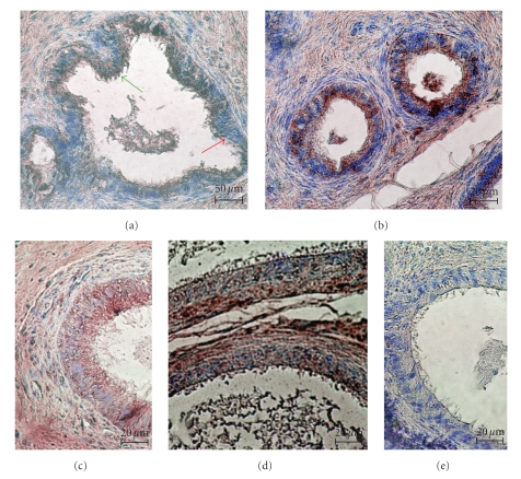
Figure 1.
The distribution of FSH-R in the ductuli efferentes (a) and in the ductus epididymis (b–d) of rat. (a) Immunostaining of FSH-R in the apical cytoplasm of nonciliated cells (red arrow) and ciliated cells (green arrow) of ductuli efferentes. (b) The strong intensity of immunoreactions in all principal cells of the caput epididymis. (c-d) The decreased staining in the principal cells of the corpus (c) and the cauda (d) epididymis. (e) The lack of immunostaining in cells of the epididymal epithelium in negative control of reaction with omitting of primary antibody. Scale bar: (a) 50 μm, (b–e) 20 μm.

