
Figure 2.
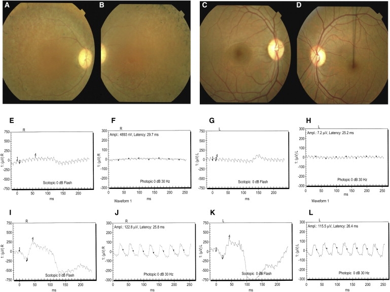
Fundus Photographs and Electroretinography Responses Illustrating the Retinal Phenotype of Family PKRP179
(A–D) Fundus photographs of individual V:5 Oculus Dexter (OD) (A) and Oculus Sinister (OS) (B) and individual V:6 OD (C) and OS (D). Individual V:5 shows several features associated with RP, including a waxy pallor of the optic disc, attenuated arterioles, atrophy of RPE, and peripheral bone spicules, which are absent in unaffected individual V:6.
(E–L) Electroretinography responses of individual V:5 are OD combined rod and cone response (E), OD cone response (F), OS combined rod and cone response (G), and OS cone response (H); and of individual V:6 are OD combined rod and cone response (I), OD cone response (J), OS combined rod and cone response (K) and OS cone response (L). Individual V:5 has typical RP changes, including loss of both rod and cone responses (E–H), whereas ERG readings of unaffected individual V:6 show no changes in the rod and cone response (I–L).