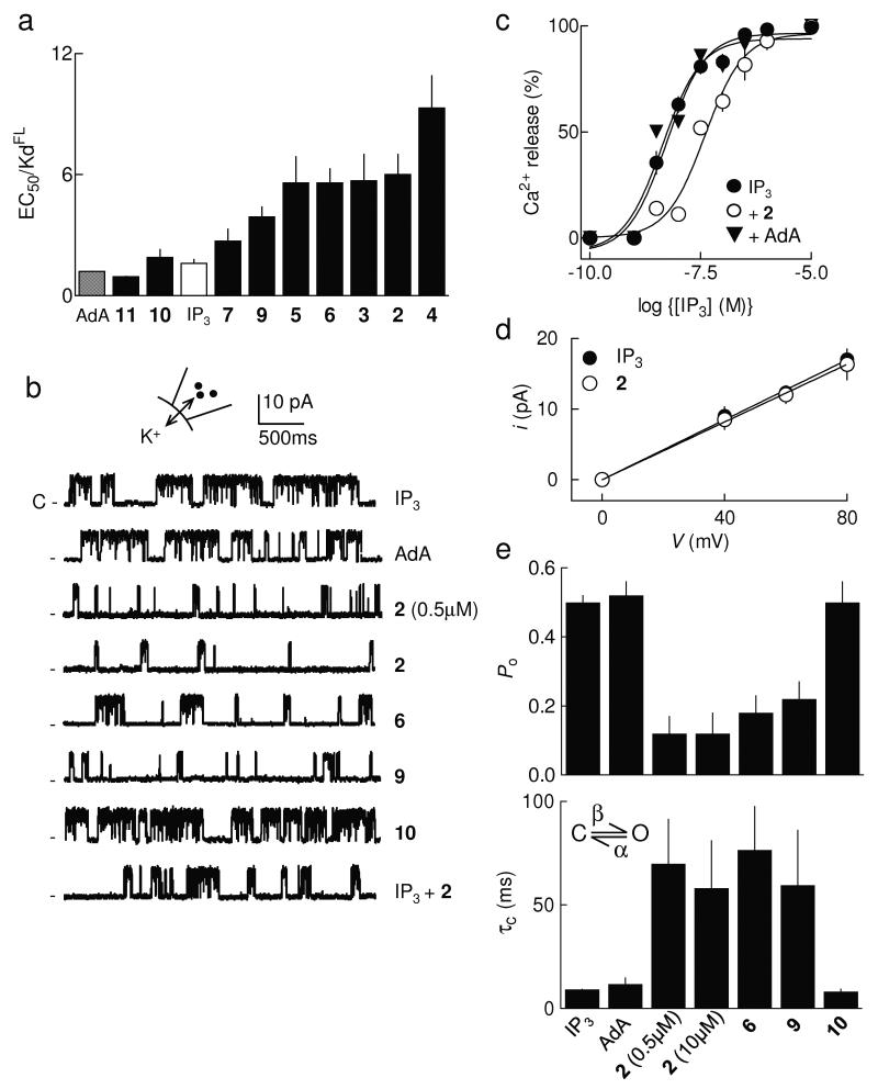
Figure 2.
2-O-modified IP3 analogues are partial agonists of IP3R. (a) From experiments similar to those shown in Supplementary Fig. 1a,b online, EC50/Kd ratios (n ≥ 4) were calculated for each ligand. (b) Traces (each typical of at least 3 similar records) show excised nuclear patch-clamp recordings from DT40-IP3R1 cells with the pipette solution containing ATP (0.5mM), a free [Ca2+] of 200nM and the indicated ligands (10μM, except where shown otherwise). The holding potential was +40mV. C denotes the closed state. (c) Cells were treated with IP3 alone, or for 30s with either 0.1nM AdA or 2 and then with the indicated concentrations of IP3. Results (n = 3) show the concentration-dependent release of Ca2+ by IP3. (d) Current-voltage (i-V) relationship for patches stimulated with IP3 or 2 (means ± SEM, n = 3). (e) Summary data showing Po and mean closed time (τc) for IP3R1 stimulated as shown, n = 3-11 (further details in Supplementary Table 1 online). The simplified activation scheme for IP3R shows the transition between closed (C) and open (O) states determined by rate constants, β and α (see Supplementary Methods online). All results (a,c-e) are means ± SEM.

