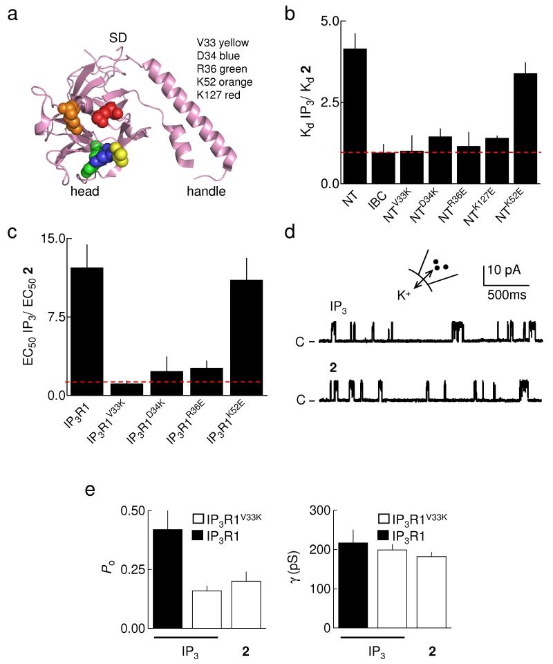
Figure 4.
Point mutations within the SD mimic partial agonists. (a) The structure of the SD33 is shown highlighting the residues mutated in this study. (b) Relative affinities (Kd) of IP3 and 2 for IBC, NT, and NT with the indicated mutations (Supplementary Table 3 online); n ≥ 5. The dashed line shows KdIP3/Kd2 = 1. (c) Potency (EC50) of IP3 relative to 2 in releasing Ca2+ from permeabilized DT40 cells stably expressing mutant IP3R1 (Supplementary Table 4 online); n ≥ 5. The dashed line shows EC50IP3/EC502 = 1. (d) Typical recordings from excised nuclear patches of DT40-IP3R1V33K cells with 10μM IP3 or 2 in the patch pipette. The holding potential was +40mV. C denotes the closed state. (e) Summary data showing Po and γK for IP3R1 and IP3R1V33K stimulated with 10μM IP3 or 2; n ≥ 3. Results (b,c,e) are means ± SEM.

