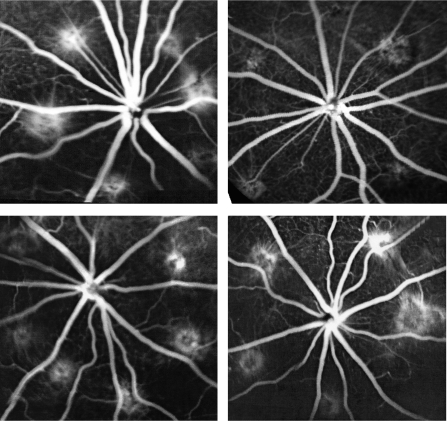
Figure 4.
Fluorescein angiographs (FA) taken in a late stage (5 minutes) at 2 weeks after laser coagulation. Top left: data from the left eye, which received 3 μL of 5% dextrose; top right: data from the fellow eye (right eye), which received 3 μg HDP-P-AraG 3 days before laser. Compared with the left eye (six laser burns), the right eye (five laser burns) showed less hyperfluorescein staining of the laser burns with minimal leakage. Bottom left: left eye, which that received 3 μL of 5% dextrose; bottom right: the fellow eye (right eye), which received 3 μg of AraG 3 days before laser photocoagulation. FA leakage from the laser burns on both eyes was comparable (six burns in the left eye and five burns in the right eye).

