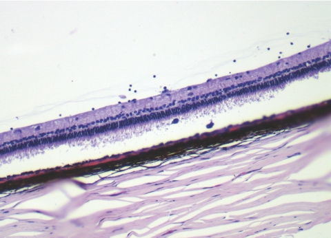
Figure 6.
A light micrograph of an eye with a 350-μg intravitreal injection of HDP-cP-5-F-2dUrd at 5 months after the injection, showing mild inflammatory cells in the inferior vitreous at the site of the drug depot and two pigment-laden macrophages in the corresponding subretinal space. The retinal detachment in the image was an artifact from the histology processing. Magnification, ×25.

