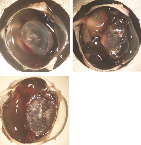
Figure 7.
Top left: an eye that had PVR-induced trauma along with intravitreal injection of 700 μg HDP-cP-5-F-2dUrd, showing a normal medullary ray and attached retina at 3 weeks. Top right: an eye that had the trauma procedure but with an intravitreal injection of 5% dextrose, showing a proliferating tissue band between the lens and medullary ray (large arrow) and detached medullary ray and retina. The retina was also pulled to the incision site (small arrow). Bottom: an eye that had PVR-induced trauma and intravitreal injection of 700 μg 5-FU, showing a proliferating stiff strand (small arrow) between the incision and the optic nerve head along with the detached medullary ray and the surrounding retina (large arrow). Magnification, ×12.

