Figure 5.
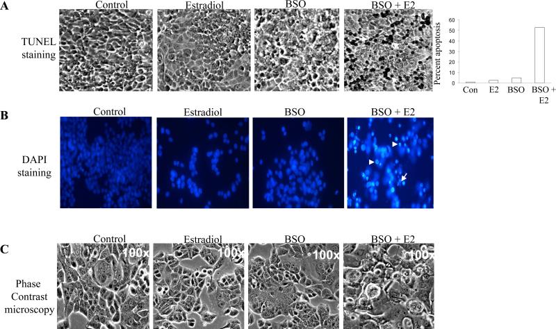
BSO enhances the apoptotic effect of estradiol in MCF-7:2A breast cancer cells. (A) Cells were treated with 1 nM E2, 100 μM BSO, or 1 nM E2 + 100 μM BSO for 72 hours and TUNEL staining for apoptosis was performed as described in “Materials and methods.” Slides were photographed through brightfield microscope under 100X magnification. TUNEL-positive cells were stained black (white arrows). Columns (right), mean percentage of apoptotic cells (annexin V-positive cells) from three independent experiments done in triplicate; bars, SEs. (B). Fluorescent microscopic analysis of apoptotic cells stained with 4',6-diamidino-2-phenylindole (DAPI). MCF-7:2A cells were treated with 1 nM E2, 100 μM BSO, or 1 nM E2 + 100 μM BSO as described above for 72 hours. To assess the number of cells undergoing apoptosis, round and/or shrunken nuclei of DAPI-stained cells were counted (white arrows). At least 200 cells per slide were counted by two individuals to control for subjective variability. Experiments were repeated three times with similar results. Representative slides are shown. Scale bars = 50 μm. (C) Phase contrast microscopy of MCF-7:2A cells treated with 1 nM E2, 100 μM BSO, or 1 nM E2 + 100 μM BSO for 72 hours.

