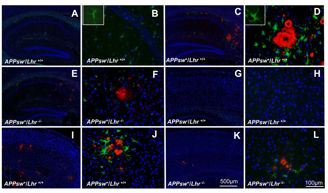
Figure 2.
Double-immunofluorescent labeling of Aβ to show amyloid plaques (red) and glial fibrillary acidic protein (GFAP) to detect astrocytes (green) in the hippocampus (A–F) and cerebral cortex (G–L) of female mice with the different indicated genotypes. DAPI staining (blue) labels cell nuclei. The inset in panel B is a normal astrocyte at higher magnification. The inset in panel D is a reactive astrocyte at higher magnification that displays thick processes and enlarged cell bodies. Scale bars: A, C, E, G, I, K = 500 µm; B, D, F, H, J, L = 100 µm.

