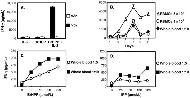
Figure 3.
Interferon (IFN)–γ secretion in response to phosphoantigens by peripheral-blood mononuclear cells (PBMCs) and whole blood. A, Dependence on γδ T cells of IFN-γ secretion in response to bromohydrin pyrophosphate (BrHPP). γδ T cells were depleted from PBMCs by positive selection with antibody-coated magnetic beads. γδ T cell–depleted and undepleted PBMCs (2 × 10⁶) were stimulated with BrHPP, interleukin (IL)–2, or BrHPP plus IL-2. Culture supernatants were collected after 7 days, and IFN-γ was measured by ELISA. One representative experiment of 3 is shown. B, Comparison of IFN-γ secretion by PBMCs and whole-blood cultures after stimulation with BrHPP. PBMCs (1 × 10⁵ and 3 × 10⁵ cells/well) and whole blood (1 mL of 1:10-diluted peripheral blood) from the same subject were stimulated in 24-well plates with BrHPP (50 μmol/L) and IL-2 (25 U/mL), and culture supernatants were harvested at indicated time points. IFN-γ was measured by ELISA. Results are expressed as the IFN-γ produced in response to BrHPP plus IL-2 minus the IFN-γ produced in response to IL-2 alone, for each time point. C–D, IFN-γ secretion by whole-blood cultures in response to different concentrations of BrHPP and isopentenyl pyrophosphate (IPP). Heparinized peripheral blood (1 mL) was diluted 1:5 (open circles) or 1:10 (black squares) with RPMI 1640 and stimulated in 24-well plates with indicated concentrations of BrHPP (C) or IPP (D) plus IL-2 (25 U/mL). Culture supernatants were collected after 7 days, and IFN-γ was measured by ELISA. Results are expressed as IFN-γ in response to IL-2 only or IL-2 plus phosphoantigen.

