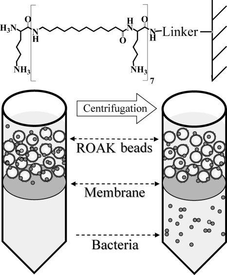
FIG. 1.
ROAK design and experimental apparatus. Shown is the molecular structure of K-7α12 linked to a polystyrene bead. Underneath is a cartoon illustration of the capture experiment: bacteria (small circles) incubated with ROAK beads (large circles) were driven to translocate across the membrane by centrifugation.

