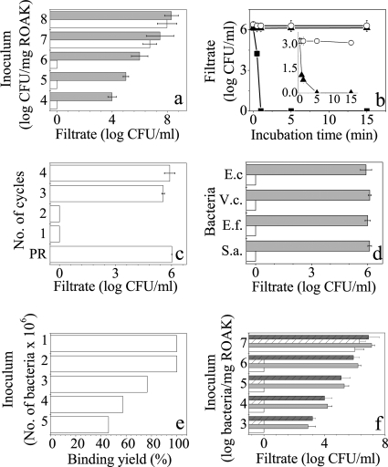
FIG. 2.
Characteristics of bacterial binding to K-7α12 ROAKs. (a) CFU count obtained from capture assay filtrates, after incubation with protected (gray bars) and deprotected (white bars) ROAKs, using the specified E. coli inocula. Error bars (a, c, d, and f) represent standard deviations from the means obtained from at least 4 independent experiments performed in duplicate. An absence of bars indicates consistency. Zero inoculum values indicate negative cultures (i.e., <10 CFU/ml). (b) Time dependence of E. coli capture. CFU count was determined in filtrates after incubation for the specified time periods with deprotected (squares) and protected (circles) ROAKs. K15 beads (triangles) were used as a control. (Inset) Low-concentration experiment comparing K15 (triangles) and the control resin (circles). (c) Depletion experiment to determine the capacity for binding to ROAK beads after repeated incubation/filtration cycles. Protected ROAKs (PR) were used for a one-cycle control experiment. (d) Assessment of binding specificity using protected (gray bars) and deprotected (white bars) ROAKs assayed as in panel a. E.c, E. coli; V.c., V. cholerae; E.f., E. faecalis; S.a., S. aureus. (e) Assessment of binding yield using direct counts of elevated numbers of bacteria before and after passage through ROAK beads. (f) Binding of bacterial communities. CFU counts from capture assay filtrates after incubation with protected (gray bars) and deprotected (white bars) ROAKs, using the specified inoculum of defined bacterial mix (E. coli, V. cholerae, E. faecalis, and S. aureus, at equal concentrations; nonstriped bars) or undefined microbial mix (treated sewage water; striped bars).

