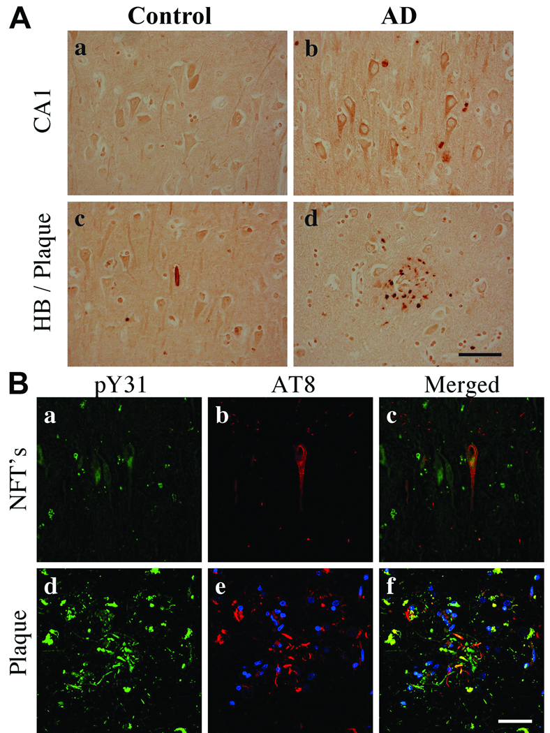
Figure 7.
Phosphorylated Y118 (pY118) paxillin in control and Alzheimer disease (AD) hippocampi. (A) CA1 pyramidal neurons exhibit nuclear pY118 in control and pY118 in granulovacuolar (GVD) bodies in AD. Insets are high power magnifications of CA1 pyramidal neurons. (b) Arrows in inset indicate GVD bodies. pY118 paxillin phospho-specific and nonspecific peptide blocking controls demonstrate antibody specificity (c, d). (B) Confocal microscopy for pY118 paxillin (green), nuclei (blue) and AT8 labeled phosphorylated Tau (pTau) (red). pY118 paxillin is colocalized in the majority of AT8+ pyramidal neurons in both neurofibrillary tangles (NFTs) and processes within plaques (a–f). Note the extensive colocalization of pY118 paxillin in nuclei of neuronal and non-neuronal cells. Scale bars: A = 50 µm for a and b; 500 µm for c and d; B = 50 µm for all panels.

