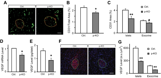
Figure 5.
Reduced islet microvascular density and lower VEGF-A expression level in STAT3 KO mice. A, Five-micrometer cryosections of p-KO and control mouse pancreas were immunolabeled with STAT3 (red) and endothelial marker CD31 (green). B, Vascular density, measured as CD31-stained area relative to the whole pancreas was reduced in p-KO (gray bar, n = 3 mice) compared with control mice (white bar, n = 3 mice). Images taken from 10 pancreatic sections of each animal were used for analysis. C, Vascular density in the islets (e.g. inside yellow boundary in A) and exocrine pancreas (e.g. outside yellow boundary in A) was similarly assessed as in B, Vascular density was significantly reduced in the islets (**, P < 0.01) and marginally reduced in the exocrine tissue (#, P = 0.06). Data are presented as means ± sem. Images taken from 10 pancreatic sections of each mouse, three mice per group, were used for analysis. D, VEGF-A mRNA levels were analyzed by quantitative real-time PCR from total RNA extracted from isolated islets. P-KO mice (gray bar, n = 4) have lower VEGF-A expression than control mice (white bar, n = 4). *, P < 0.05. E, VEGF-A content in isolated islets from p-KO mice (gray bar, n = 3) was significantly lower than that from control mice (white bar, n = 3). VEGF-A level was determined by ELISA. Data are presented as means ± sem. *, P < 0.05. F, Five-micrometer cryosections of p-KO and control mouse pancreas were immunolabeled with VEGF (red) and 4′,6′-diamino-2-phenylindole (blue). Green boundary denotes islets. G, VEGF-A levels in the islets and exocrine pancreas were measured by integrating the gray levels inside and outside the islets. VEGF-A levels in the islets and exocrine pancreas decreased significantly in p-KO mice. **, P < 0.01. Multiple images were taken from pancreatic sections of each mouse, and three mice per group were used for analysis.

