
Figure 4.

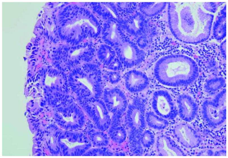
Image of high-grade dysplasia. A. Endoscopic microscope image of high grade dysplasia stained with 0.05% acriflavine. Confluent and haphazard glandular proliferation with back–to-back arrangements and minimal to absent stroma. Foci of high nuclear intensity and high nuclear–to-cytoplasmic ratio are highlighted by markers (red arrowheads). B. Histopathology of same specimen. Scale bar is 100 microns; all images are at the same scale.