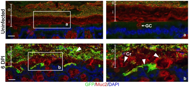
Figure 1. Citrobacter rodentium penetrates the colonic mucus layer in vivo.
Staining for GFP-expressing C. rodentium using an antibody that recognizes GFP (green), and murine Muc2 (red), with DAPI (blue) as a counterstain. No GFP-labeled C. rodentium can be seen in the mucus layers of uninfected mice (upper panels), but in infected mice, C rodentium is observed within the outer and inner mucus layer in regions where the underlying epithelium is infected (bottom panels). Right panels “a” and “b” are expanded images of corresponding boxed regions in left panels. o = outer mucus layer; i = inner mucus layer; Cr = C. rodentium; GC = goblet cell. Original magnification = 200×. Scale bar = 50 µm.

