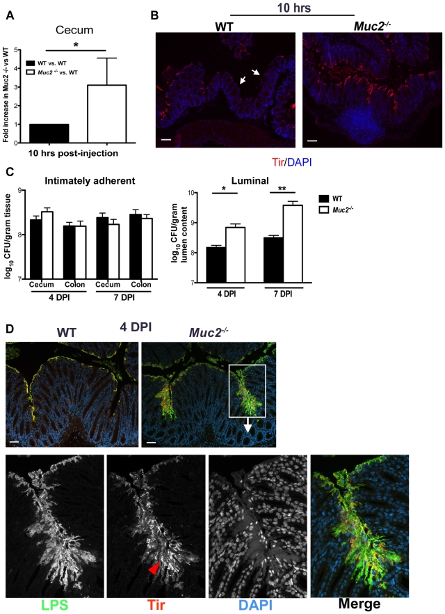
Figure 5. Muc2 limits initial colonization of the mucosal epithelia, but ultimately controls levels of luminal pathogen burdens.
A. Fold differences of intimately adherent C. rodentium numbers present in the ceca of WT vs Muc2−/− mice 10 hours post-injection of 1.5×10⁸ CFU into cecal lumen in a cecal loop surgery experiment (see Material & Methods). Results are of data from a total of 5 mice per group pooled from 2 individual experiments. Error bars = SEM (*P = 0.0109, Mann-Whitney test). B. Representative immunostaining for the C. rodentium-specific effector Tir in ceca acquired from cecal loop surgery, 10 hrs post-injection. C. rodentium is found on the surface of Muc2−/− cecal mucosa in a continuous fashion compared to WT mice, where Tir staining is patchy amid long stretches of uncolonized surface epithelium (white arrows). Original magnification, 100×. Scale bar = 100 µm. C. Quantification of luminal C. rodentium vs. intimately adherent C. rodentium attached to the cecal and colonic mucosa in WT vs. Muc2−/− mice at 4 and 7 DPI. Results represent the mean value pooled from 2 independent infections containing 3–4 mice per group. Error bars = SEM (*P = 0.0140; **P = 0.005, Mann-Whitney test). D. Visualization of C. rodentium infection by staining for LPS (green) and Tir (red; red arrowhead), with nuclei specific DAPI (blue).as a counterstain. Tir staining is localized to the surface epithelium in both WT and Muc2−/− mice indicating direct infection, but the majority of LPS-positive cells in Muc2−/− mice are not infecting (Tir-negative), yet are accumulating on the surface of the mucosa. Original magnification, 200×. Scale bar = 50 µm.

