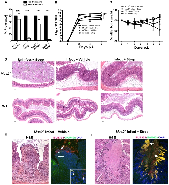
Figure 10. Antibiotic induced commensal depletion enhances pathogen colonization but does not alter host pathology in Muc2−/− mice.
A. Quantification of DAPI stained bacteria from stools of WT and Muc2−/− mice 24 hours following oral treatment with Streptomycin (20 mg) or Vehicle (dH20). Streptomycin (strep) led to significantly reduced numbers of total bacteria within mouse stool. Results represent the means of 3–4 mice per group. Error bars = SEM (***P<0.001, unpaired t test). B. Enumeration of ΔespF C. rodentiumStr (strep-resistant) in stool of strep-or vehicle-treated mice as indicated, at various times post-infection. Results represent the means of 3–4 mice per group. Error bars = SEM (*P≤0.05, Mann-Whitney test, one-tailed). C. Body weights following infection of strep or vehicle treated WT and Muc2−/− mice with ΔespF C. rodentiumStr. n = 3–4 mice per group. Error bars = SEM. D. Representative histological sections of ceca from uninfected or infected (6 DPI) strep- or vehicle-treated WT and Muc2−/− mice. Original magnification = 100×. Scale bar = 100 µm. E. H&E (Left panel) and FISH analysis (right panel) of an ulcer from ΔespF C. rodentiumStr infected vehicle-treated Muc2−/− mouse cecum (6 DPI). Numerous commensals (EUB338+/GAM42− cells, red) can be seen overlying the ulcer in direct contact with PMNs (arrow), and both pathogen (EUB338+/GAM42a+ cells, yellow) and commensal (red) can be seen within the PMNs (arrow heads, inset). Original magnification = 200×. Scale bars = 100 µm. F. H&E and FISH analysis of an ulcer in the descending colon from an ΔespF C. rodentiumStr infected strep-treated Muc2 −/− mouse (6 DPI). Large pathogenic microcolonies (yellow) are associated with the ulcer (arrows), while commensals (red) can be seen in the lumen. Original magnification = 200×. Scale bar = 100 µm.

