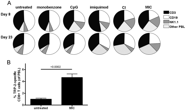
Figure 4. MIC-treated mice show a sustained NK cell expansion and circulating melanocyte antigen-specific CD8+ T cells.
A, Peripheral blood was collected from the tailvein of treated mice on day 8 and 23 of treatment, and average ratios between T cells (CD3+, black sections), B cells (CD19+, white sections), NK cells (NK1.1+, CD3-, grey sections) and other peripheral blood leukocytes (PBL; dashed sections) were determined for the PBL (n = 7 mice per group). Interestingly, MIC-treated mice showed a significant NK cell expansion on both day 8 and 23 (see table 2, “Exp. 4”). This expansion of NK cells was also found in CpG-, imiquimod- and CI-treated mice, although for these animals this reaction was only found on day 8. Monobenzone alone did not influence PBL ratios on either time point, comparable to untreated mice. Depicted data is representative of three independent in vivo mouse experiments. For the statistical analysis of the in vivo differences in NK cell counts in these experiments see table 2. B, Peripheral blood CD8+ T cells were tested for binding to H2-Kb/TRP-2180-188-tetramers at day 120 following tumor inoculation (day 85 after treatment cessation, n = 4). TRP-2 represents one of the immunodominant epitopes of B16.F10 melanoma. Long-term surviving, MIC-treated mice showed a significant population of TRP-2-specific CD8+ T cells circulating in their peripheral blood at day 120, as compared to untreated mice 10 days after tumor inoculation (n = 7). Binding to control H2-Kb/OVA257-264-tetramer by the tested PBL was negative (data not shown).

