Figure 7.
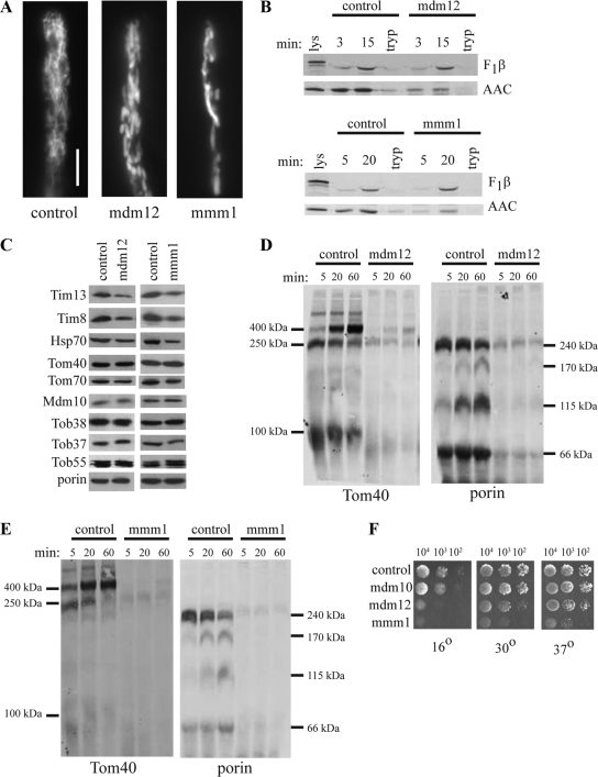
Characterization of mitochondria from mdm12 and mmm1 mutants. (A) Hyphae were incubated with MitoTracker Green and examined by fluorescence microscopy. Bar, 10 μm. (B) Precursors of F1β and AAC were imported into mitochondria isolated from the mdm12 and mmm1 mutant strains as described in Figure 2. (C) Mitochondrial proteins in the mdm12 and mmm1 mutants. Mitochondria were isolated from the indicated strains and subjected to Western blot analysis (30 μg mitochondrial protein per lane) using antibodies directed against the indicated proteins. (D) Import of radiolabeled Tom40 (left) and porin (right) into mitochondria isolated from the mdm12 mutant as described in the legend to Figure 2. (E) Import of Tom40 (left) and porin (right) into mitochondria isolated from the mmm1 mutant as described in the legend to Figure 2. (F) Growth rate of mutant strains. Conidiaspores from the control (76-26), and indicated mutant strains were counted and diluted to the desired concentrations. 104, 103, and 102 conidia from each strain were spotted on plates containing Vogel's medium with sorbose. The plates were incubated at 16°C for 72 h, 30°C for 48 h, or 37°C for 48 h and then photographed.

