Figure 6.
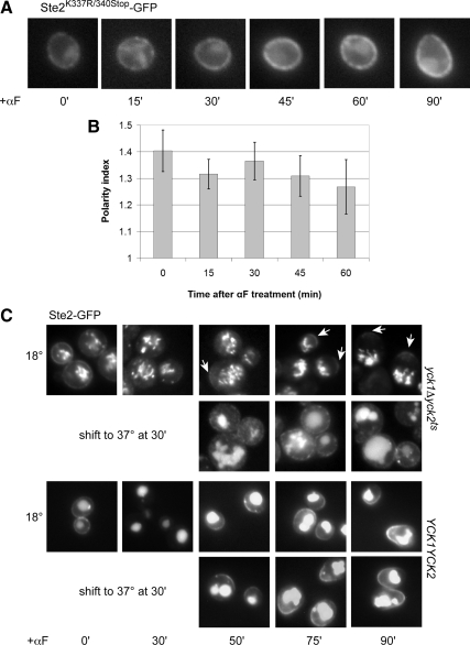
Pheromone-induced polarization of the receptor requires its internalization. (A) Representative fluorescent images of Ste2K337R/340Stop-GFP localization. G1-synchronized cells were treated with 30 nM pheromone (0 time), and images were acquired every 15 min. (B) Quantification of Ste2K337R/340Stop-GFP polarization. The bar graphs represent the mean polarity index ± SD at each time point (n = 15). (C) Representative fluorescent images of Ste2-GFP localization in yck1Δ yck2ts mutant and YCK1 YCK2 isogenic control strains. Cells were grown to mid-log phase at 18°C and treated with 600 nM pheromone (0 time) for 30 min to allow for the initial internalization of the receptor. Half the culture was then shifted to the restrictive temperature (37°C), and images were acquired at the indicated time points. Arrows mark the Ste2-GFP crescents. None of the mutant cells cultured at restrictive temperature displayed detectable crescents (n = 66 at 75 min; n = 84 at 90 min), whereas polarized localization of Ste2-GFP was clearly visible in about a third of the yck1Δ yck2ts cells cultured at the permissive temperature (33%, n = 106 at 75 min; 31%, n = 101 at 90 min).

