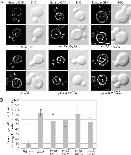
Figure 3.
Loss of the known nuclear targets of Slt2p, Rlm1p, and Swi4p or of the cell wall stress sensors, Wsc1p and Mid2p, has no significant effect on the ER distribution in ptc1Δ cells. (A) Representative fluorescence of the ER marker Hmg1-GFP and DIC images of wild-type, ptc1Δ, ptc1Δ rlm1Δ, ptc1Δ swi4Δ, ptc1Δ wsc1Δ, and ptc1Δ mid2Δ cells grown to early log phase in SC medium at 25°C. Open arrowheads point to small buds. Bars, 2 μm. (B) The ER distribution in small buds (0.3–0.5 diameter of mother cell) of all six strains shown in A was quantified. Error bars, SEM from three independent experiments.

