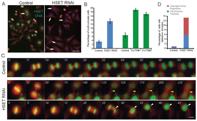
Figure 1. HSET Perturbation Causes Polyploid Cell Formation.
(A) HeLa cells were transfected with Luciferase RNAi oligo (Control) or HSET RNAi oligo, fixed, and stained for MTs (red), HSET (green), and DNA (blue). Arrows indicate multi-nucleate cells. Scale bar = 50 μm. (B) Mean percentages of multi-nucleate cells in control and HSET RNAi cells reported as mean ± SEM, p<0.01. The right half of the graph (green) shows a separate negative control oligo and two additional HSET oligos. (C) Live imaging of HeLa cells expressing GFP-H2B and mCherry-tubulin in control (top row, Movie S1) and HSET (middle and bottom rows, Movies S2 and S3) RNAi cells. Middle row, HSET RNAi movie showing ‘cleavage furrow regression’ defect, indicated by the yellow arrowhead (Movie S2). Bottom row, HSET RNAi movie depicting the ‘traveling chromosomes’ phenotype, indicated by the white arrowhead (Movie S3). Scale bar = 10 μm. (D) Total percentages of phenotypes described in part C.

