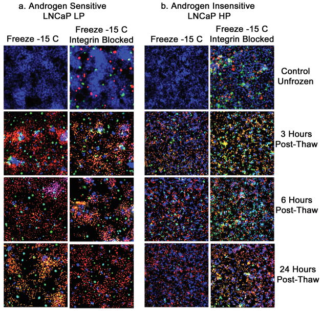
Figure 4. Fluorescent micrographs of AS and AI prostate cancer cells following freeze exposure with and without integrin inhibition.
Total levels of necrotic and apoptotic cell death were evaluated in LNCaP LP (A) and LNCaP HP (B) cells treated with 40μg/ml anti-α6β4 integrin antibody. Samples were frozen at −15°C and triple-probe fluorescent micrographs were taken after 3h, 6h, and 24h using Hoechst (blue) to assess viable cells, propidium iodide (red) to assess necrotic cells, and YO-PRO-1 (green) to assess apoptotic cells. A) Compared to freeze alone, AS cell line LNCaP LP with function-blocked integrins showed only slight increases in necrotic and apoptotic cells. B) Function blocked LNCaP HP exhibited significant increases (p < 0.05) in necrotic and apoptotic cell death at every tested time point, indicating that AI cell lines more greatly depended on integrin signaling pathways for increased freezing resistance.

